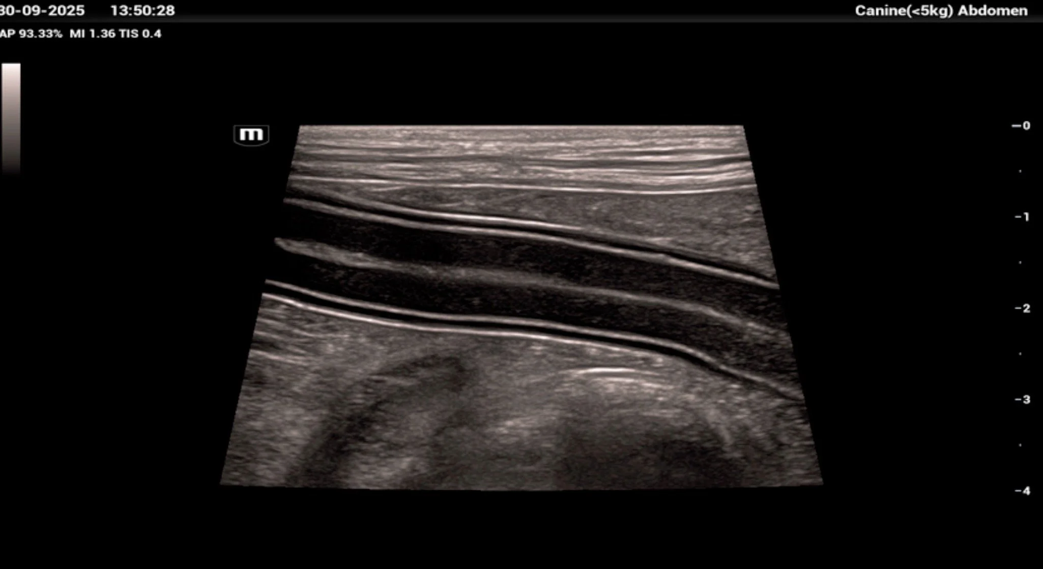
High-resolution ultrasonographic image of canine jejunum demonstrating all five histological layers: mucosal interface, mucosa, submucosa, muscularis externa, and serosa. The clarity of stratification supports normal architecture and provides a baseline for comparison in cases of enteritis, lymphangiectasia, or neoplasia.

Histological Layering of the Duodenum, Jejunum, and Ileum in Dogs and Cats: A Comparative Guide

The small intestine of dogs and cats—comprising the duodenum, jejunum, and ileum—shares a conserved four-layered histological architecture: mucosa, submucosa, muscularis externa, and serosa. Despite this overarching similarity, each segment exhibits distinct structural and functional adaptations that reflect its role in digestion, absorption, and immunological surveillance. Understanding these differences is essential for interpreting diagnostic imaging, histopathology, and surgical anatomy in clinical practice.

Mucosa

The mucosa is the innermost layer and consists of three subcomponents: the epithelium, lamina propria, and muscularis mucosae. In all three intestinal segments, the epithelium is composed of simple columnar cells with abundant microvilli forming the brush border, interspersed with goblet cells that increase in density distally. The lamina propria is a loose connective tissue matrix housing capillaries, lymphatics, and immune cells. The muscularis mucosae is a thin layer of smooth muscle that facilitates local mucosal movement.

In the duodenum, the mucosa is characterized by tall, leaf-like villi and deep crypts of Lieberkühn. The villi are more robust proximally, reflecting the duodenum’s role in nutrient emulsification and early absorption. The jejunum displays long, finger-like villi with a high surface area-to-volume ratio, optimized for maximal nutrient absorption. In contrast, the ileum exhibits shorter, blunter villi and a higher density of goblet cells, consistent with its role in water and electrolyte absorption and mucosal protection.

Submucosa

The submucosa is a dense connective tissue layer containing larger blood vessels, lymphatics, and the submucosal (Meissner’s) plexus. In the duodenum, the submucosa is notable for the presence of Brunner’s glands, which secrete alkaline mucus to buffer gastric acid. These glands are absent in the jejunum and ileum. The ileum’s submucosa is distinguished by prominent aggregated lymphoid nodules—Peyer’s patches—which are critical for mucosal immunity. These patches are more numerous and organized in the ileum than in the jejunum or duodenum, and their presence can be appreciated grossly and histologically.

Muscularis Externa

This layer consists of two smooth muscle layers: an inner circular and an outer longitudinal layer, separated by the myenteric (Auerbach’s) plexus. This arrangement is consistent across all three segments and is responsible for peristalsis and segmental contractions. In dogs and cats, the muscularis externa is relatively uniform, although subtle regional differences in thickness and innervation density may exist. The jejunum, for example, may exhibit slightly more prominent segmentation activity, reflected in the rhythmic contraction patterns observed during ultrasonography.

Serosa

The outermost layer is the serosa, a thin layer of connective tissue covered by mesothelium. It provides a frictionless interface with surrounding organs and is consistent in appearance across the small intestine. In surgical and imaging contexts, the serosa is critical for assessing intestinal integrity and identifying pathological adhesions or perforations.

Comparative Notes and Clinical Relevance

While the layering is conserved between dogs and cats, feline intestines tend to be slightly narrower and more delicate, with proportionally longer villi and a higher goblet cell density. These differences may influence the interpretation of biopsy samples and the response to enteric pathogens. For example, feline enterocytes exhibit a higher turnover rate, which may contribute to the species’ sensitivity to certain chemotherapeutic agents and toxins.

In contrast, pathological states such as lymphangiectasia, inflammatory bowel disease, or neoplasia disrupt this layering—flattening villi, distorting crypts, and infiltrating the lamina propria—changes that can be correlated with ultrasonographic and endoscopic findings.

References

Evans HE, de Lahunta A. Miller’s Anatomy of the Dog. 4th ed. Elsevier; 2013.

Dellmann HD, Eurell JA. Textbook of Veterinary Histology. 6th ed. Wiley-Blackwell; 1998.

Jubb KVF, Kennedy PC, Palmer N. Pathology of Domestic Animals. Vol 2. Academic Press; 2007.

Washabau RJ, Day MJ. Canine and Feline Gastroenterology. Elsevier; 2012.

Pereira MA et al. Histological and morphometric evaluation of the small intestine of dogs. Acta Scientiae Veterinariae. 2016;44(1):1–8.