Ultrasonographic Evaluation of Liver Masses in Dogs: Diagnostic Capabilities and Limitations
QUIZ
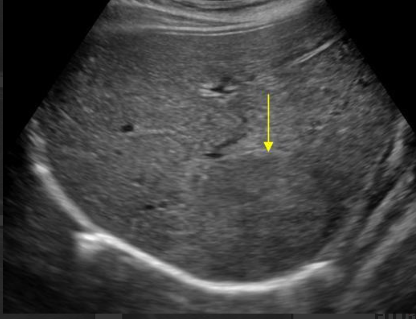
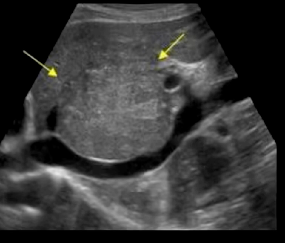
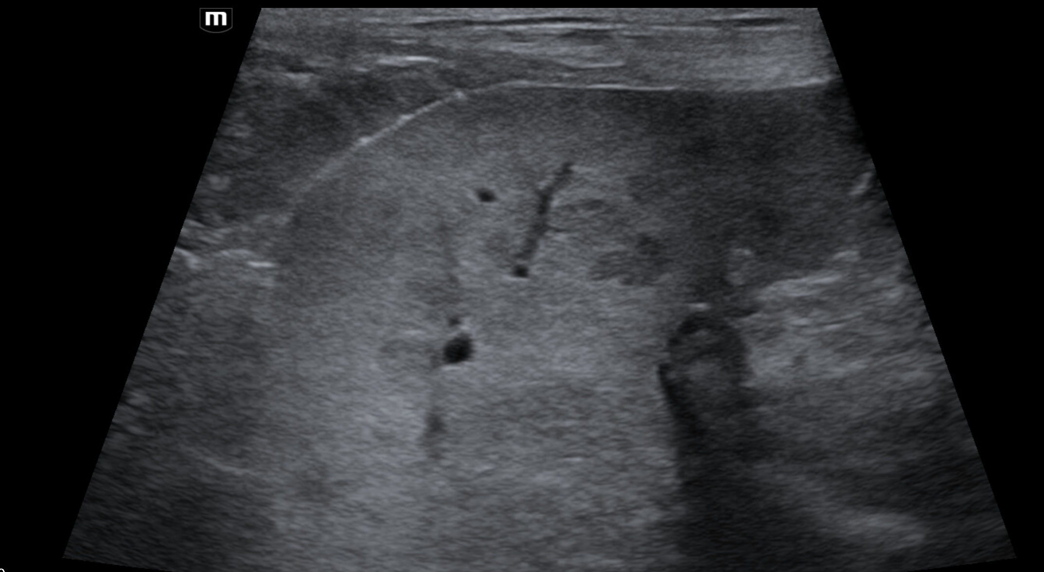
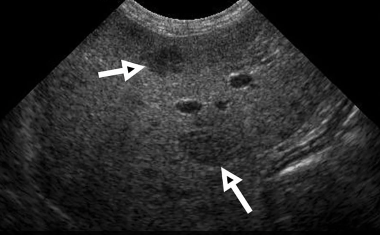
Which of these liver masses/nodules is a malignant tumor.
Focal liver lesions are a common ultrasonographic finding in dogs, particularly in middle-aged to older patients. These lesions encompass a wide spectrum of pathological processes, including benign hyperplastic nodules, primary hepatic neoplasia, and metastatic disease. Ultrasonography is the first-line imaging modality for hepatic assessment due to its availability, lack of ionizing radiation, and ability to guide sampling. However, despite continual advances in ultrasound technology, imaging characteristics alone are frequently insufficient to differentiate benign from malignant liver masses, making cytologic or histopathologic sampling essential in the majority of cases.
This article reviews the ultrasonographic appearance of canine liver masses, including conventional B-mode imaging, Doppler, contrast-enhanced ultrasound (CEUS), and elastography, with emphasis on diagnostic accuracy, reported statistics, and the critical role of tissue sampling.
Prevalence and Classification of Canine Liver Masses
Primary hepatic tumors are uncommon in dogs, accounting for approximately 0.6–1.5% of all canine neoplasms (Patnaik et al., 1980; Liptak et al., 2004). Among primary hepatic neoplasms:
Hepatocellular carcinoma (HCC) represents approximately 35–60%
Cholangiocarcinoma (biliary carcinoma) accounts for 22–41%
Sarcomas (e.g. hemangiosarcoma) and neuroendocrine tumors are rare
(Liptak et al., 2004; Withrow & Vail, 2019)
In contrast, metastatic disease is significantly more common, with hepatic metastases occurring 2.5–3 times more frequently than primary liver tumors (Nyland et al., 2002; d’Anjou & Penninck, 2015). Common primary sources include splenic hemangiosarcoma, intestinal carcinoma, pancreatic carcinoma, mast cell tumor, and lymphoma.
Benign lesions are also frequent, particularly in geriatric dogs. Nodular hepatocellular hyperplasia is extremely common, with post-mortem studies demonstrating microscopic or macroscopic nodules in up to 70–100% of dogs over 10–14 years of age (Johnson et al., 1995; Cullen & Stalker, 2016).
B-Mode Ultrasonography
Detection and Morphology
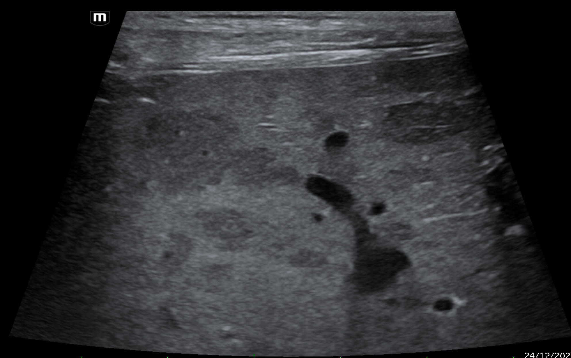
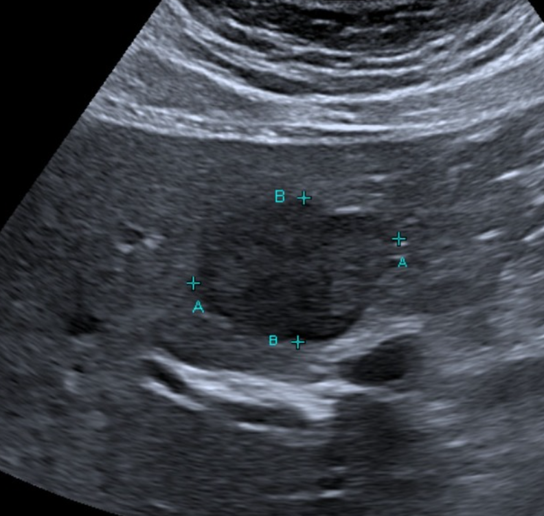
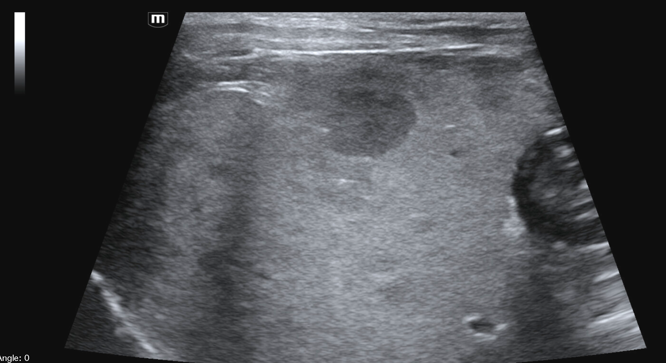
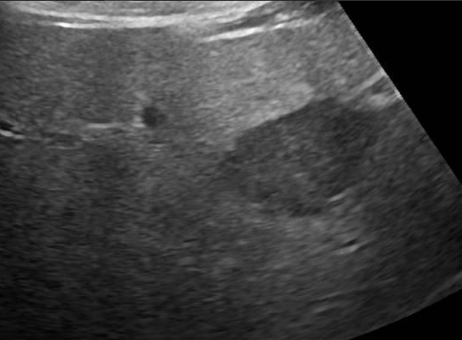
B-mode ultrasonography is highly sensitive for detecting focal liver lesions but is poorly specific for lesion type. Hepatic masses may appear:
Hypoechoic, hyperechoic, or mixed echogenicity
Well-marginated or irregular
Solitary or multifocal
Importantly, echogenicity does not correlate reliably with malignancy. Multiple studies have demonstrated substantial overlap in the sonographic appearance of benign nodules, primary hepatic tumors, and metastatic lesions (Nyland et al., 2002; O’Brien et al., 2004)
In a large retrospective study of canine liver tumors, no consistent B-mode ultrasonographic features reliably differentiated hepatocellular carcinoma, cholangiocarcinoma, sarcoma, or metastatic lesions, aside from lesion distribution (O’Brien et al., 2004). Hepatocellular carcinomas were more likely to be solitary, whereas sarcomas and metastases were more often multifocal, but significant overlap existed.
Sensitivity for Metastatic Disease
While ultrasound is commonly used for staging, its sensitivity for detecting hepatic metastases is limited. For example:
In dogs with splenic hemangiosarcoma, ultrasonographic detection of hepatic metastases had a sensitivity of approximately 19%, despite a specificity of ~82% (O’Brien et al., 2004).
In dogs with mast cell tumors, sensitivity for hepatic metastasis was reported at 29%, with specificity exceeding 90% (Stefanello et al., 2009).
These findings emphasize that a normal hepatic ultrasound does not exclude metastatic disease, and conversely, that many ultrasonographically visible nodules are benign.
Doppler Ultrasonography
Color and power Doppler ultrasound allow assessment of vascular architecture and blood flow within hepatic lesions. Malignant tumors may demonstrate
Internal or peripheral neovascularization
Tortuous or irregular vessel
However, Doppler findings overlap considerably between benign and malignant lesions, and vascularity alone cannot be used to determine tumor type (d’Anjou & Penninck, 2015). Doppler imaging is most useful for:
Identifying major vessels prior to biopsy
Avoiding hemorrhagic complications
Supporting, but not confirming, malignancy
Enhancement Patterns
CEUS provides dynamic assessment of lesion perfusion during arterial and portal phases using microbubble contrast agents. Several enhancement trends have been described:
Hepatocellular carcinoma: commonly shows arterial-phase hyperenhancement followed by portal-phase washout
Cholangiocarcinoma: variable enhancement, often iso- or hypoenhancing
Sarcomas: frequently show minimal or absent enhancement due to necrosis
Metastases: often demonstrate rapid arterial enhancement with early washout
(Lassau et al., 2011; Zini et al., 2013; Bargellini et al., 2016)
In a large retrospective study evaluating CEUS in canine liver tumors, arterial hyperenhancement was observed in approximately 74% of hepatocellular carcinomas, while most sarcomas failed to enhance (Zini et al., 2013).
Diagnostic Limitations
Despite these trends, significant overlap exists, and CEUS cannot definitively distinguish benign from malignant lesions. Zini et al. (2013) concluded that although CEUS improves lesion characterization, histopathology remains mandatory for diagnosis, as no enhancement pattern was pathognomonic.
Elastography
Ultrasound elastography assesses tissue stiffness and is an emerging modality in veterinary imaging. Preliminary studies suggest:
Malignant liver lesions tend to be stiffer than benign nodules
Benign hyperplastic nodules are generally softer